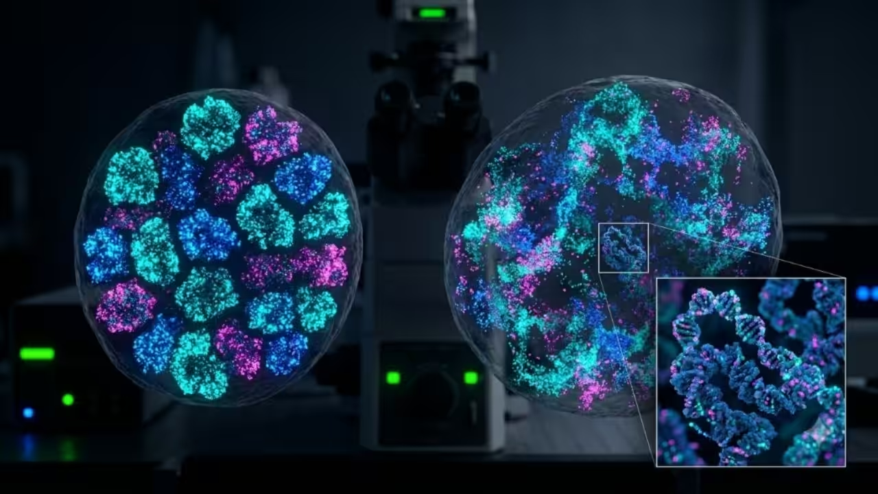
Scientists have developed a new class of fluorescent probes capable of showing how DNA is packaged and moves inside living cells at remarkably fine scales. The technology produced images of chromatin in living cells at about 20-nanometer resolution and located individual dye molecules in preserved samples with around three-nanometer precision.
The research involved scientists from the Center for Genomic Regulation in Barcelona, the City University of Hong Kong, and the Guangdong Provincial People’s Hospital. The findings were published in the peer-reviewed journal Molecular Cell.
Table of Contents
DNA Packaging Helps Control Gene Activity

A human cell contains roughly two meters of DNA, yet this material must fit inside a nucleus measuring only a few micrometers across. Cells accomplish this feat by wrapping DNA around proteins and organizing it into chromatin.
This packaging is not merely a space-saving arrangement. It helps determine which genes can be accessed by the machinery that reads genetic information. Loosely packed DNA is generally more accessible, while tightly folded regions tend to be less active.
Chromatin also changes as cells grow, divide, respond to stress, or develop specialized functions. Disruptions to this organization have been associated with cancer and other diseases, making DNA folding an important area of biological and medical research.
New HoT Probes Blink Automatically
The newly developed fluorescent molecules are known as HoT probes. They are Hoechst–TAMRA derivatives that combine a DNA-binding Hoechst component with a rhodamine-based fluorescent structure.
After entering a cell, the probes attach to DNA and spontaneously switch between bright and dark states. This blinking behavior allows microscopes to separate fluorescent signals that would otherwise overlap.
If all the probes emitted light simultaneously, their signals would create a blurred image. Because individual molecules blink at different times, researchers can record their positions across thousands of frames. Computer software then combines those observations into a detailed reconstruction.
The probes are also permeable to living cells and sufficiently photostable for extended observations. Technical details of their design and testing are available in the official study published in Molecular Cell.
Living DNA Viewed at 20-Nanometer Resolution
Researchers tested the probes in living human skin cells and laboratory-grown HeLa cancer cells. They combined the dyes with stochastic optical reconstruction microscopy (STORM) to examine chromatin at a resolution of approximately 20 nanometers.
Conventional optical microscopes cannot clearly resolve structures smaller than about 200 nanometers due to the diffraction limit of light. STORM works around that barrier by calculating the locations of fluorescent molecules that appear separately over time.
The resulting images were approximately ten times sharper than those produced by conventional light microscopy. More importantly, scientists were able to watch DNA move inside living cells rather than studying only chemically fixed snapshots.
This could help researchers track chromatin as cells change identity, respond to environmental signals, or develop abnormal characteristics.
Preserved Cells Delivered Near-Molecular Precision
The team obtained its most precise measurements from preserved laboratory cells, in which cellular structures were fixed in position. Using an advanced microscopy method known as MINFLUX, the scientists located individual fluorescent molecules with approximately three-nanometer precision.
The DNA double helix is about two nanometers wide, placing the measurement close to the molecule’s physical scale. However, localization precision should not be confused with the overall image resolution. The result means that the position of a single dye molecule could be estimated within roughly three nanometres; it does not mean every feature of the DNA strand was directly resolved at that scale.
The researchers have made supporting microscopy material available through the study’s official imaging dataset, which covers live cells, preserved cells and tissue sections.
Cancer Cells Showed Looser DNA Folding
To explore the method’s medical potential, researchers examined wax-preserved bowel tissue obtained from three cancer patients. Hospitals and pathology laboratories routinely store this type of preserved biopsy material.
The images showed that chromatin in the tumor cells appeared looser and more dispersed than in nearby healthy cells. Such changes are biologically significant because altered DNA packaging can affect gene activity, DNA repair, and genomic stability.
Pathologists already assess changes in the size, shape, and appearance of cell nuclei when examining suspected cancers. Measuring how DNA occupies three-dimensional space could eventually provide another layer of information.
The result remains preliminary, however. Samples from three patients cannot establish a diagnostic test or show whether chromatin looseness reliably predicts a tumor’s aggressiveness. Validation would require much larger studies involving multiple cancer types and disease stages.
Artificial Intelligence Recognized Cell Types
The researchers also analyzed the new images using AINU, which stands for “AI of the Nucleus.” The deep-learning system was introduced in 2024 to identify subtle patterns in super-resolution images of cell nuclei.
After training on images collected from living cells, AINU distinguished skin cells from stem cells with an accuracy of between 96% and 98%. Both cell types contain essentially the same DNA sequence, but they organize and use their genetic material in different ways. The system was therefore detecting structural patterns connected to cellular identity.
The Center for Genomic Regulation previously explained how AINU identifies nanoscale changes inside cell nuclei. Combining the software with live-cell probes enables scientists to explore whether those patterns change over time.
Potential applications include identifying promising stem cells for regenerative research and distinguishing abnormal cells from healthy ones, although these possibilities have not yet been demonstrated in clinical settings.
Zebrafish Tests Could Support Regeneration Research
The probes also worked in sections of zebrafish eye tissue. Zebrafish can regenerate damaged retinal cells far more effectively than humans, making them useful models for studying tissue repair.
Regeneration is thought to require some cells to become more flexible and to reactivate genetic programs that are normally silent. This process may involve loosening chromatin to allow previously inaccessible genes to be expressed again.
By measuring DNA compaction in individual retinal cells, the new system could help researchers investigate how genome organization changes during repair. The probes do not stimulate regeneration themselves; they provide a more detailed method for observing the cellular events associated with it.
A Clearer View of the Genome’s Hidden Structure
The technology still has important limitations. HoT probes label DNA broadly rather than a particular gene, and the most precise measurements were obtained from preserved cells rather than living ones. STORM and MINFLUX equipment also remain highly specialized.
Even so, the method brings together live tracking, nanoscale imaging, tissue compatibility, and artificial-intelligence analysis within a single experimental platform. The Centre for Genomic Regulation’s official research report describes possible future uses ranging from cancer research to retinal regeneration.
For now, the probes are research tools rather than medical products. Their immediate value lies in helping scientists study the genome as a moving, three-dimensional structure whose physical arrangement can influence how cells behave in health and disease.