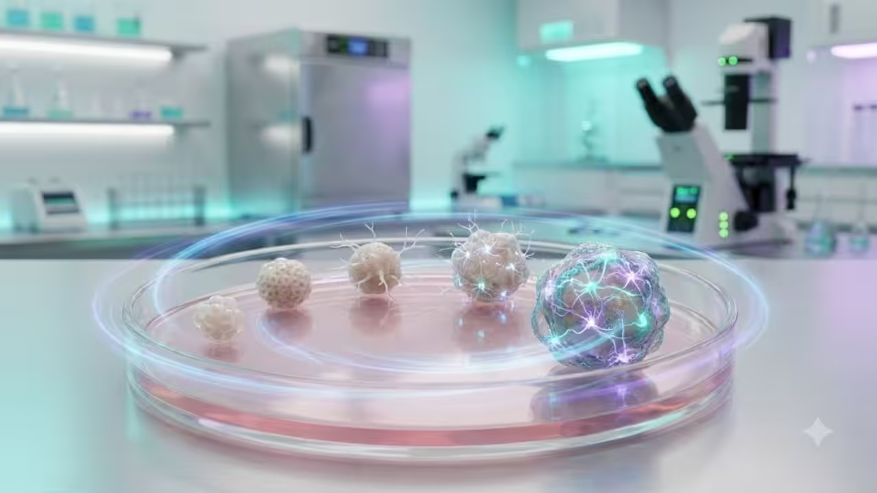
Tiny clusters of human brain cells, known as brain organoids, have not only survived but also thrived in a Harvard laboratory for over five years. This remarkable duration gives scientists a unique chance to study how the human cerebral cortex matures in a controlled environment, far longer than in previous studies.
These organoids, roughly the size of a peppercorn, have demonstrated significant developmental progress. Their cells have undergone various molecular and epigenetic transformations that mirror the natural progression of brain development, including changes typically observed after birth. This research is particularly promising, as it lays the groundwork for a valuable platform to explore neurological disorders, such as autism and schizophrenia, and to test potential treatments in a setting that closely resembles human brain development.
The longevity of these brain organoids allows researchers to study the intricate processes involved in cortical maturation over an extended period, offering insights into how neural circuits form and how they may be affected by genetic and environmental factors. This could lead to breakthroughs in understanding the complexities of brain development and the underlying mechanisms of various neurological conditions.
Table of Contents
Five-Year Cultures Set a New Longevity Record
Scientists have previously maintained human cortical organoids for extended periods, but the longest reported lifespan was 694 days. That benchmark came from a 2021 study on long-term cortical organoid maturation, which found that the tissue acquired characteristics resembling early postnatal development.
The Harvard team has now extended that timeline beyond five years, roughly three times the earlier record. Some related cultures in the laboratory have reportedly reached seven years, although the published analysis primarily covers development through five years.
According to Harvard’s official account of the research, the organoids were small enough to resemble peppercorns but contained well over one million cerebral cortex cells each.
How Scientists Created the Brain Organoids
The organoids were derived from cells collected from human donors. Researchers reprogrammed these cells into induced pluripotent stem cells, which can develop into many specialised cell types. Carefully timed biochemical signals then directed them towards the formation of neurons, neural progenitor cells, astrocytes, and other cells found in the cerebral cortex.
This process produces a three-dimensional model that captures selected features of developing brain tissue. It allows scientists to observe cellular events that would be difficult or ethically impossible to study directly inside a living human brain.
However, the structures are not miniature human brains. They lack blood vessels, sensory input, a complete anatomical organisation, and connections to the rest of the body. They model particular aspects of cortical development rather than replicating the entire brain.
Nearly 425,000 Cells Tracked Over Time
The researchers examined 34 organoids at eight stages between six months and five years. They used single-cell RNA sequencing to identify which genes were active inside individual cells at each stage.
After combining the findings with previously collected data, the team assembled information from 110 organoids across 16 time points, ranging from 15 days to five years. The complete analysis covered 424,720 cells.
Early organoids between 15 days and two months displayed gene-expression patterns resembling first-trimester human brain development. Cultures between three and six months more closely matched the second trimester. From nine months onwards, the cells gradually developed molecular signatures resembling those of late prenatal and postnatal stages.
An Internal Clock Recorded the Passage of Time
A central question was whether the organoids were genuinely developing or merely surviving. The results indicated that their cells continued to move through recognisable biological stages.
One of the clearest signs came from DNA methylation, a process that adds chemical markers to DNA and helps regulate whether particular genes are active. Methylation patterns change in a broadly predictable way with age, allowing researchers to use them as biological clocks.
The team analysed whole-genome methylation at nine time points between three months and five years. The organoids accumulated age-related changes that broadly paralleled those seen in human cortical tissue.
Researchers identified 213 DNA regions whose methylation differed between three-month-old and five-year-old organoids. Many were linked to genes and regulatory elements involved in the formation of the cerebral cortex. Two established methylation clocks also showed strong correlations between the predicted biological age and the actual time spent in culture.
Older Cells Showed a Developmental Memory
The study’s most intriguing experiment involved mixing young and old neural progenitor cells in chimeric organoids, sometimes called “chimeroids.” These structures contained cells from different donors and developmental stages.
After receiving signals that encouraged neurone formation, young progenitors produced early-stage cell types. Nine-month-old progenitors exposed to the same environment behaved differently: they generated later-stage neurones and glial-related cells.
Within two weeks, older progenitors produced cell types that would normally take two or three months to emerge in a young culture. The cells appeared to retain molecular information about the developmental stages they had already completed.
Researchers described the effect as a developmental “time warp”. It does not represent conscious memory. Instead, it indicates that cells can preserve a biological record of age and use that information when responding to new signals.
Modified Conditions Protected Fragile Neurones
Long-term organoid research faces a major obstacle: neurones are fragile and often decline as cultures age. Supporting cells, such as astrocytes, tend to survive more reliably.
The Harvard team tested a modified, activity-permissive growth medium containing lower glucose, physiologically adjusted ion levels and a stabilised glutamine supplement. The environment encouraged spontaneous electrical signalling and provided additional metabolic support.
Organoids grown under these conditions developed more neurons, denser synapses and more elaborate neuronal branches. By one year, they showed coordinated bursts of electrical activity that were absent in conventionally maintained cultures. Researchers continued detecting this activity for up to two years.
Electrical signalling alone does not indicate thought or consciousness. It demonstrates that neurones were communicating through functional networks within a limited laboratory model.
Findings Could Expand Neurological Research
Long-lived organoids may help scientists investigate stages of brain development that occur around birth and during early childhood. The Arlotta Laboratory at Harvard studies how cortical cell types emerge, build circuits and become affected by disease.
Organoids made from different donors could reveal how genetic variation influences development. They may eventually support research into autism, schizophrenia, epilepsy and neurodegenerative conditions, while also providing human-cell models for evaluating experimental medicines.
The study did not produce a treatment or demonstrate that organoid tissue can be safely transplanted. Its immediate contribution is a richer experimental model of development.
A Longer Window Into Human Brain Development
Important limitations remain. Organoids lack vascular and immune systems, and cells in their cores may experience oxygen and nutrient shortages. Maintaining cultures for several years is also expensive, technically demanding and impractical for routine experiments.
The next objective is therefore not simply to keep organoids alive longer. Researchers want to identify the mechanisms controlling their developmental clock and reproduce later stages more quickly.
By showing that cortical organoids can survive, mature and retain their biological age over several years, the work provides a longer opportunity to study human brain development, one that scientists previously had no practical way to observe in the laboratory.